
Clinical Acupuncture for Stress-Induced Insomnia: Neurobiological Mechanisms and Integrative Protocols
Clinical Focus: Neuro-targeted acupuncture point selection stimulates peripheral nerve pathways to downregulate the sympathetic nervous system and induce deep, restorative sleep states.
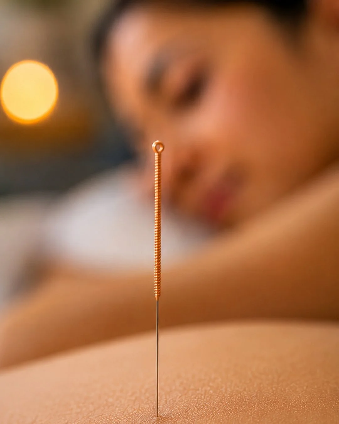
Acupuncture for stress-induced insomnia involves the strategic insertion of sterile, micro-fine needles into specific anatomical points to regulate the autonomic nervous system and restore physiological sleep cycles. In our Langley and Surrey clinics, patients experiencing chronic sleep disturbances driven by hyperarousal utilize this evidence-informed therapy to downregulate sympathetic ("fight-or-flight") dominance and stimulate parasympathetic activity.
By targeting precise neurovascular nodes, acupuncture triggers the release of endogenous opioids, serotonin, and gamma-aminobutyric acid (GABA). This biochemical shift modulates the hypothalamic-pituitary-adrenal (HPA) axis, directly reducing elevated cortisol levels that disrupt circadian rhythms. Patients undergo a thorough clinical assessment mapping their stress-response patterns, followed by targeted needling sessions tailored to their physical presentation. This safe, drug-free intervention addresses the underlying neurobiological mechanisms of sleep onset and maintenance delays, facilitating sustained, restorative rest.
The Neurobiology of Stress-Induced Insomnia
When chronic stress originates from demanding work weeks commuting along Hwy 1 or the daily pressures facing families in Willoughby and Cloverdale, the body's physiological threat-response system remains chronically active. This state of hyperarousal alters the central nervous system, creating a distinct physical presentation.
The Anatomy of Hyperarousal
In a normal circadian cycle, cortisol levels drop in the evening, allowing melatonin to rise. For patients with stress-induced insomnia, this rhythm is fractured. The HPA axis remains reactive, keeping the sympathetic nervous system engaged long after sunset (Pilozzi et al., 2020).
Physically, this manifests as:
Persistent Muscular Hypertonicity: The suboccipital muscles at the base of the skull, the upper trapezius, and the levator scapulae feel dense, fibrous, and locked in a defensive shrug.
Elevated Core Metrics: Mild tachycardia (elevated resting heart rate), shallow thoracic breathing rather than deep diaphragmatic expansion, and vasomotor instability.
Neurological Restlessness: An overstimulated cortex leads to involuntary nocturnal behaviors—frequent position shifting, jaw clenching (bruxism) that strains the temporomandibular joint, and hyper-awareness of environmental sounds.
How Acupuncture Directs Sleep Neurophysiology
Acupuncture does not act as a sedative; rather, it functions as a biological modulator that balances autonomic nervous system activity (Spence, 2004). The insertion of a 0.25mm stainless steel needle into cutaneous and muscular tissue initiates a cascade of localized and systemic neurological events.
1. Neurotransmitter Regulation
The mechanical stimulation of peripheral nerve fibers transmits signals to the spinal cord, midbrain, and hypothalamus (Bae et al., 2025). Clinical data indicates this stimulation prompts the brain to increase the synthesis of GABA and endogenous melatonin (Spence, 2004). GABA reduces neuronal excitability throughout the nervous system, effectively quieting the "racing mind" described by patients lying awake in Brookswood or Murrayville.
2. HPA Axis De-escalation
By modulating the limbic system and the hypothalamus, acupuncture inhibits the overproduction of Corticotropin-Releasing Hormone (CRH) (Bae et al., 2025). As a result, systemic cortisol levels drop. When blood cortisol decreases, the physiological barriers to sleep onset dissolve, allowing the brainstem to transition naturally into slow-wave sleep.
3. Local Tissue Decompression
Stress-induced insomnia rarely exists without physical discomfort. Tight, ischemic muscles restrict local capillary blood flow, sending pain signals back to the brain, which acts as a secondary wakefulness trigger. Acupuncture induces local vasodilation, flushing metabolic waste from hypertonic tissues in the neck, shoulders, and back, terminating the pain-insomnia feedback loop.
Clinical Protocols and Point Selection
Our integrative approach focuses strictly on evidence-based point selections that correlate with identified neuroanatomical structures.
Anmian (Extra Point): Located posterior to the cranium, midway between Yifeng (TE17) and Fengchi (GB20). It targets the lesser occipital nerve and occipital artery. It is deeply sedating and specifically reduces sleep-onset latency and suboccipital tension.
Yintang (Extra Point): Located at the midpoint between the medial extremities of the eyebrows. It stimulates branches of the trigeminal nerve. It downregulates frontal lobe hyperarousal and is clinically observed to halt repetitive, anxious thoughts.
Shenmen (HT7): Located at the ulnar end of the transverse crease of the wrist. It sits adjacent to the ulnar nerve and artery to modulate autonomic tone, stabilize heart rate variability (HRV), and mitigate nocturnal anxiety.
Sanyinjiao (SP6): Located on the medial aspect of the lower leg, 3 cun superior to the medial malleolus. It sits near the posterior tibial nerve and is clinically associated with improved sleep quality metrics and endocrine balance.
During a session in our quiet, clinical environment, a patient from Walnut Grove or Langley City will lie in a recumbent position. Once the needles are placed, a distinct sensation known as De Qi is typically achieved—characterized by a deep, dull ache, heavy warmth, or a radiating tingle. This sensation confirms the targeted nerve pathways have been successfully engaged.
The needles remain in place for 20 to 30 minutes, during which time objective shifts in autonomic function can often be observed: respiration slows, the pulse softens, and borborygmus (stomach growling) occurs, signaling a successful shift into parasympathetic digestion and rest states.
The Integrative Advantage at Near Me Therapy
Insomnia is multifactorial. Because our clinic houses Registered Acupuncturists alongside Chiropractors, RMTs, Kinesiologists, and Counsellors, we do not view sleep disruption in isolation.
For instance, a patient whose insomnia is exacerbated by chronic lower back pain or postural strain from long desk hours in Willoughby may receive acupuncture to regulate their nervous system, while concurrently working with our RMTs to release structural myofascial restrictions. If somatic anxiety is a primary driver, clinical counselling works in tandem with acupuncture to provide cognitive behavioral tools alongside somatic down-regulation (Liu et al., 2021).
We track progress objectively, monitoring markers such as:
Reduction in sleep onset latency (time taken to fall asleep) (Spence, 2004).
Decreased nocturnal awakenings and improved sleep efficiency (Kalavapalli & Singareddy, 2007).
Reduction in self-reported daytime fatigue and jaw tension.
Improved heart rate variability (HRV) metrics where applicable.
Acupuncture offers a measurable, biologically viable pathway out of stress-induced sleep fragmentation, restoring the physical architecture necessary for your body to heal itself overnight.
References
Bae, R., Kim, H. Q., Lu, B., Ma, J., Xing, J., & Kim, H. Y. (2025). Role of Hypothalamus in Acupuncture’s Effects. Brain Sciences, 15(1), 72. https://doi.org/10.3390/brainsci15010072
Kalavapalli, R., & Singareddy, R. (2007). Role of acupuncture in the treatment of insomnia: A comprehensive review. Complementary Therapies in Clinical Practice, 13(3), 184-193. https://doi.org/10.1016/j.ctcp.2007.01.001
Liu, C., Zhao, Y., Qin, S., Wang, X., Jiang, Y., & Wu, W. (2021). Randomized controlled trial of acupuncture for anxiety and depression in patients with chronic insomnia. Annals of Translational Medicine, 9(18), 1426-1426. https://doi.org/10.21037/atm-21-3845
Pilozzi, A., Carro, C., & Huang, X. (2020). Roles of β-Endorphin in Stress, Behavior, Neuroinflammation, and Brain Energy Metabolism. International Journal of Molecular Sciences, 22(1), 338. https://doi.org/10.3390/ijms22010338
Spence, D. W. (2004). Acupuncture Increases Nocturnal Melatonin Secretion and Reduces Insomnia and Anxiety: A Preliminary Report. Journal of Neuropsychiatry, 16(1), 19-28. https://doi.org/10.1176/appi.neuropsych.16.1.19